
Breast Cancer Bone Metastasis
Context of Use or Disease: Osteolytic bone metastasis in breast cancer
Platform: Emulate - Chip-S1
Description: This model comprises a tri-culture organ-on-a-chip system incorporating murine breast cancer cells (4T1), osteoclast precursors (RAW 264.7-derived osteoclasts), and osteocytes (MLO-Y4). Osteocytes are embedded in a collagen-I matrix within one channel, while cancer cells are cultured in a parallel channel separated by a porous membrane, enabling paracrine signalling. Osteoclast differentiation is induced using M-CSF and RANKL.
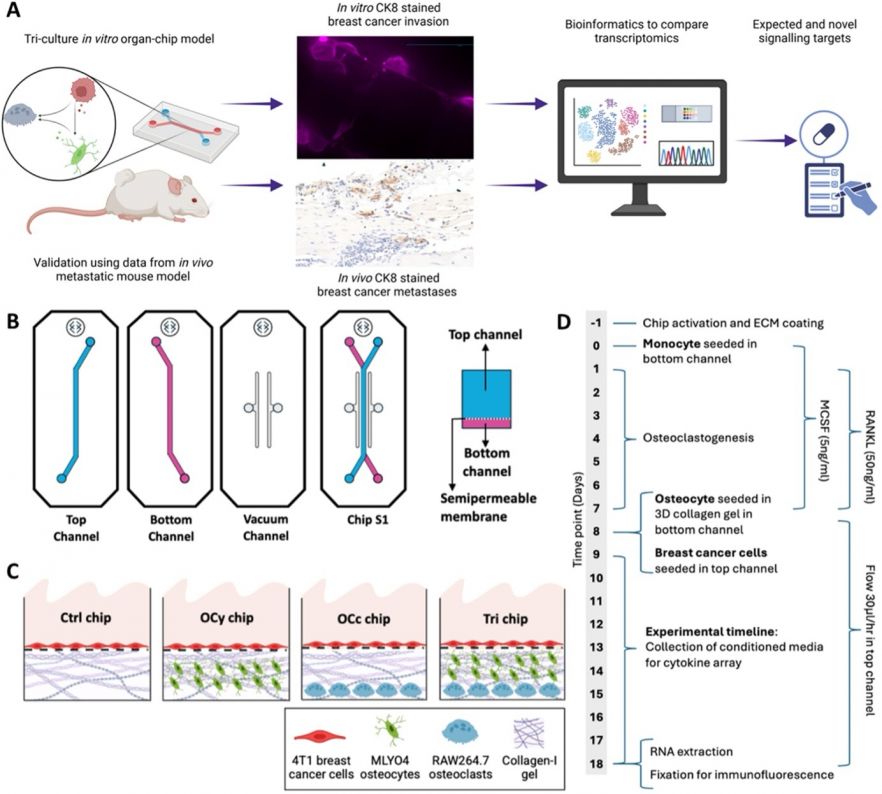
Fig. 1. Schematic showing osteolytic metastases chip layout and experimental design.
Characterisation & Validation: The model is validated using multi-omics approaches, including RNA sequencing, cytokine profiling, and fluorescence imaging. It reproduces key features of osteolytic metastasis, including increased cancer cell migration, osteoclast activity, and inflammatory signalling. Transcriptomic profiles show strong alignment with in vivo mouse models of bone metastasis, with synergistic interactions between cell types driving pro-metastatic and bone-degrading pathways. The use of multi-omics and improved RNA extraction from fixed chip samples is expected to enhance reproducibility and enable broader adoption in preclinical research.
Ongoing Research: Mechanistic understanding osteocyte-regulated tumour progression, identification of novel therapeutic targets within the metastatic niche, integration of osteoclasts.
Research Team: Natalia Muñoz Castro, Eleni Maniati, Oliver Pearce, Stefaan Verbruggen, Martin Knight
Lead Contact: Stefaan Verbruggen
Last updated 15/05/2026