
Vascularised Synovium
Context of Use or Disease: Synovial inflammation, synovitis, arthritis
DOI: Biomedical Materials 2023
Platform: Emulate - Chip-S1
Description: Healthy human fibroblast-like synoviocytes (hFLS) were cultured in the top channel, with human umbilical vein endothelial cells (HUVECs) in the bottom channel, both with appropriate matrix proteins, and separated by a flexible, porous membrane. Cyclic tensile strain was applied at 0-12% to mimic physiological activity. The pro-inflammatory cytokine, interleukin−1β (IL−1β), was added into the synovium channel and THP-1 monocytes were added to the vascular channel.
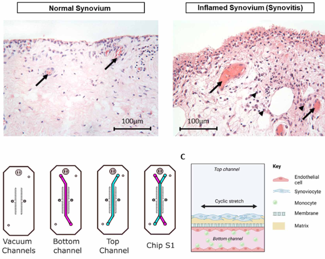
Fig. 1. Schematic showing the design of the vasculaised synovium organ-chip
Characterisation & Validation: The hFLS exhibited characteristic morphology, cytoskeletal architecture and matrix protein deposition. Stimulation with IL−1β resulted in increased secretion of the inflammatory and catabolic mediators, interleukin-6 (IL−6), prostaglandin E2 (PGE2), matrix metalloproteinase 1 (MMP−1), as well as the synovial fluid constituent protein, hyaluronan. Enhanced expression of the inflammatory marker, intercellular adhesion molecule-1 (ICAM-1), was observed in HUVECs in the vascular channel, accompanied by increased attachment of circulating monocytes.
Ongoing Research: Addition of resident immune cells, investigation into the effects of diet/glucose/insulin
Research Team: Martin Knight, Tim Hopkins
Lead Contact: Martin Knight
Last updated 30/03/2026