
Cardiac Organoid
Context of Use or Disease: Cardiomyocyte electrophysiology
DOI: Biosensors and Bioelectronics 2023
Platform: Organoid
Description: Cardiomyocytes were deposited into round bottom 96-well plates coated with polyhydroxyethylmethacrylate (polyHEMA) overnight at 60°C to avoid adhesion. Wells were seeded with 1E5 cells each (human induced pluripotent stem cell-derived cardiomyocytes or primary cardiomyocytes), incubated for 5 days in medium until formation of a cohesive organoid.
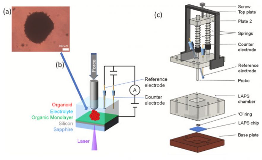
Fig. 1. Photoelectrochemical imaging of cardiac organoids pressed onto the sensor with a force probe.
Characterisation & Validation: Light-Addressable Potentiometric Sensors (LAPS) were used to evaluate the cardiac organoid electrophysiology. Cardiomyocyte contraction frequency and amplitude were measured and compared to organoids treated with phenylephrine, verapamil or blebbistatin to validate both measurement technique and cardiac organoids.
Ongoing Research: Improving the design of Light-Addressable Potentiometric Sensors, elucidating mechanisms of cardiac mechanobiology in parallel with engineered heart tissues and cardiomyocyte slice cultures
Research Team: Thomas Iskratsch, Steffi Krause
Lead Contact: Thomas Iskratsch
Last updated 28/04/2026