
Ovarian Tumour Slices
Context of Use or Disease: Ovarian cancer immune microenvironments
DOI: STAR Protocols 2024, iScience 2023
Platform: Ex vivo tissue slices
Description: Fresh human and mouse malignant ovarian cancer tissues are sliced on a vibratome and analysed by spinning disc microscopy, among other methods, to measure T cell and macrophage motility. Tumour slices can be maintained 24h ex vivo, retaining viability and cell movements.
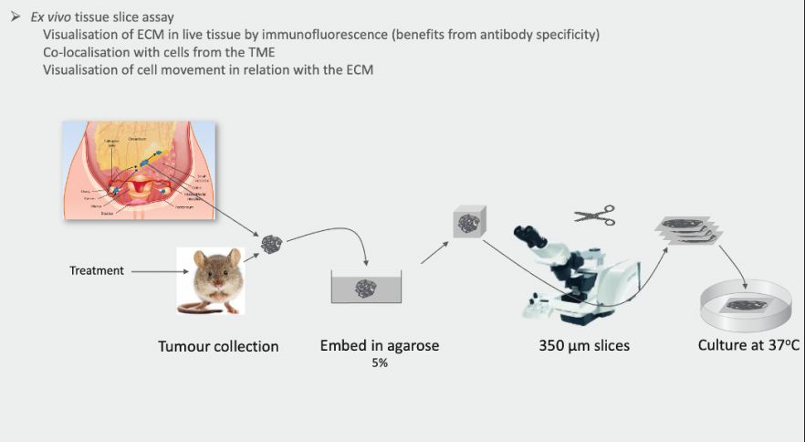
Fig. 1. Ex vivo tissue slice assay.
Characterisation & Validation: T cell and macrophage motility were characterised in live human and mouse ovarian cancer tissue slices. CD8+ T and myeloid cells can display four different behaviours ex vivo (static, wobbling, migrating or long migrating), stable for 24h and altered by lipopolysaccharide (LPS). In vivo chemotherapy leads to reduced CD8+ T and myeloid cell motility ex vivo.
Ongoing Research: Influence of therapies on T cell and macrophage motility, localisation and function in human tumours, extension of model to other normal and malignant human tissues
Research Team: Frances Balkwill, Beatrice Malacrida, Joash Joy, Panoraria Kotantaki, Eleni Maniati, Rachel Bryan-Ravenscroft
Lead Contact: Frances Balkwill
Last updated 28/04/2026