
Vascularised Ovarian Cancer
Context of Use or Disease: Omental metastasis of high grade serous ovarian cancer (HGSOC), chimeric antigen receptor (CAR)-T cell therapy in solid tumours
DOI: Cancer Research 2024
Platform: Photolithography moulded microfluidic chip
Description: The microfluidic chip consists of three parallel channels, with the central channel having a 2 mm-diameter well in the middle. Fibrin gel with human endothelial cells (ECs) is initially injected into the central channel. Established collagen gels containing human HGSOC cell lines, primary human omental fibroblasts and ECs, were then introduced into the well in the central channel. The device is cultured for 7 days, after which human CAR-T cells are introduced through the side channels and cultured for an additional 3 days.
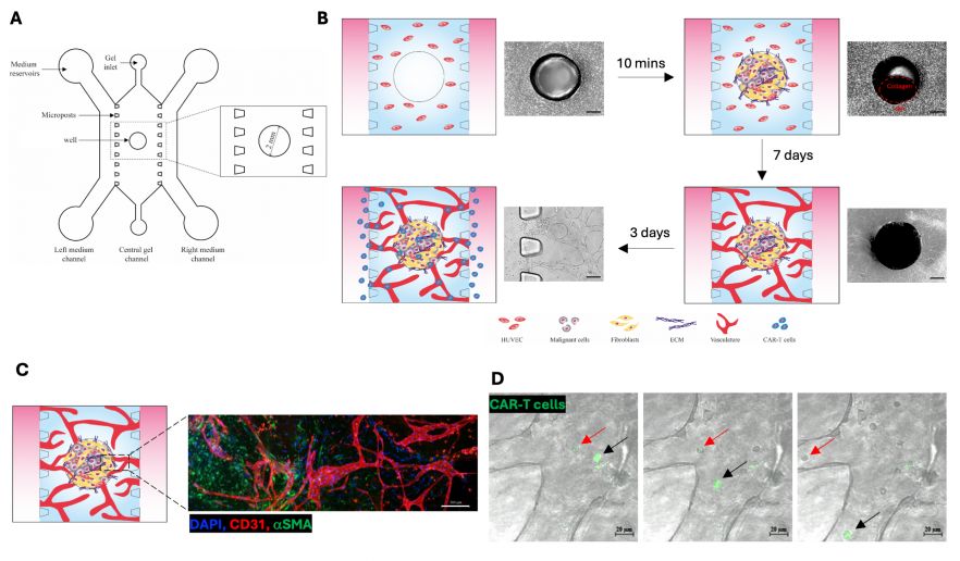
Fig. 1. Vascularised ovarian cancer-on-a-chip. (A) Design of the tri-channel microfluidic device with a 2 mm-diameter well in the central channel. (B) Schematic diagram showing model development. Scale bar: 600 μm. (C) Immunofluorescence image showing ovarian cancer on-a-chip. Cancer cells and fibroblasts are stained with alpha smooth muscle actin (αSMA; Green) and microvasculature is stained with CD31 (Red). Scale bar: 200 μm. (D) Real-time images showing the luminal flow of CAR-T cells (arrows) through the vasculature formed within the microfluidic device. Green: CAR-T cells. Scale bar: 20 μm.
Characterisation & Validation: The microvasculature was characterised by immunofluorescent staining for basement membrane and junction proteins. Immunohistochemical staining for malignant cell, fibroblast and extracellular matrix markers on the collagen gels validated features of human HSGOC. Real-time imaging confirmed the flow of CAR-T cells through the microvasculature. CAR-T cell activity was validated by staining for cell death markers and analysis of effluent for proinflammatory cytokines using Mesoscale Discovery (MSD).
Ongoing Research: Incorporation of additional cell types (primary adipocytes, mesothelial cells isolated from macroscopically normal omentum, monocytes from blood) to better study CAR-T cell activity in HGSOC omental metastasis
Research Team: Frances Balkwill, Beatrice Malacrida, Joash Joy, Panoraia Kotantaki, Eleni Maniati, Rachel Bryan-Ravenscroft, Julien Gautrot
Lead Contact: Frances Balkwill
Last updated 28/04/2026