
Intestine
Context of Use or Disease: Inflammatory bowel disease (IBD), Crohn’s disease (CD), ulcerative colitis (UC), fungal infection, tissue fibrosis
DOI: bioRxiv 2025
Platform: 3D printed microfluidic chip on microscope slides
Description: Fibrin hydrogel-based 3D organotypic human intestine model with closed loop vascular flow system. Incorporates Caco-2 gut epithelial cells, CCD-18Co fibroblasts, HUVEC vascular endothelial cells, and primary blood monocytes (or THP-1 cell line). A silicon frame is extrusion printed onto a microscope slide and used to hold a fibrin hydrogel that contains fibroblasts +/- monocytes. This forms the extracellular matrix, and a readily accessible vascular channel is created during this process. Intestinal epithelial cells are seeded onto the surface of the fibrin ECM, and HUVECs via the vascular channel.
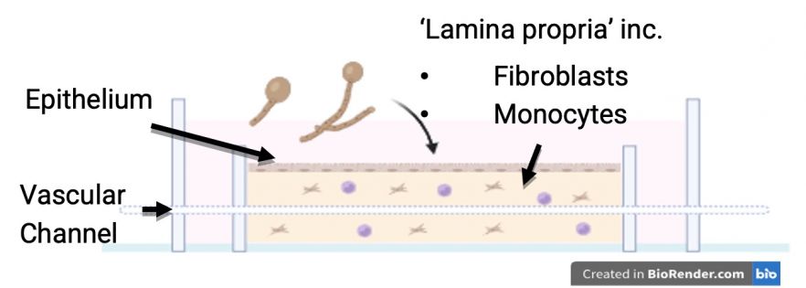
Fig. 1. Schematic showing intestine model design.
Characterisation & Validation: The intestinal models are cultured in a custom complete media for up to 7 days. Cellular morphology and cell specific markers (E-Cadherin, ZO-1, Vimentin) have been validated through H&E and immunofluorescent staining. Intact barrier function was shown using FITC-dextran paracellular permeability assays. Intestinal microbiota has been inoculated onto the epithelial surface (C. albicans). Supernatant multiplex ELISA demonstrates relevant cell-cell interaction between monocytes and fibroblasts through IL-8, IL-6 and CCL2 cytokines.
Ongoing Research: Investigation of how human intestinal fungi trigger inflammation and gut tissue remodelling / fibrosis in the context of IBD, CD and UC
Research Team: Neil McCarthy, Sean Carlson
Lead Contact: Neil McCarthy
Last updated 17/04/2026