
Microvascularised Tissue-on-Chip
Context of Use or Disease: Interfacing spheroid or 3D hydrogel-based tissue models with microvascularised networks
DOI: Scientific Reports 2024, Scientific Reports 2023, Cancer Research 2024, Frontiers in Bioengineering and Biotechnology 2022
Platform: 3D printed microfluidic chips in multi-well plate format
Description: The model is based on microfluidic chips in which spheroids, organoids, tumoroids and other 3D tissue models can be introduced via a central well, directly above a microvascularised channel, in contact with adjacent media channels. This enables mimicking the systemic delivery of molecular therapeutics, nanomaterials and cells. This was demonstrated with cardiac and ovarian cancer spheroids, and full thickness skin equivalents.
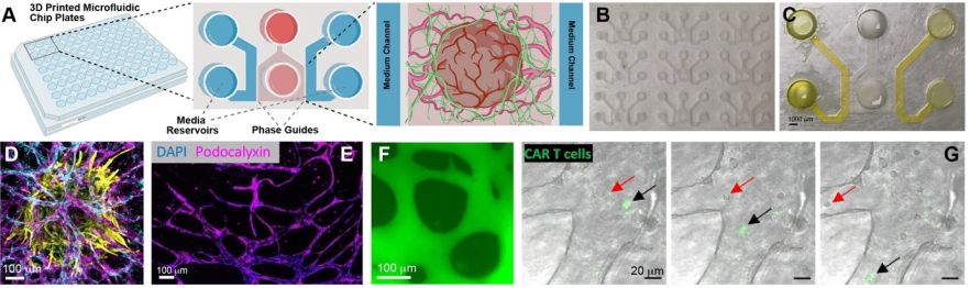
Fig. 1. A. Schematic representation of microvascularised chips with embedded ovarian cancer model. B-C. Images from a microfluidic chip plate. D. Example of spheroid (cardiac in this case; cTnT, yellow; NG2, cyan; CD31, magenta) embedded in microvascularised chips (stable up to 4 weeks of culture). E-F. Confirmation of network maturity (E, podocalyxin staining; F, FITC-dextran perfusion assay). G. CAR T cells (green) perfusing within microvascularised ovarian cancer models.
Characterisation & Validation: The stability of this model was validated, up to 4 weeks of culture. The maturity of the microvascular networks and their perfusability with macromolecules, nanoparticles and cells was demonstrated. The use of the model to investigate cardiotoxicity (in combination with cardiac spheroids) and CAR T cell therapy (for ovarian cancer) was validated.
Ongoing Research: Investigation of cardiac toxicity, testing of ovarian cancer therapies, application to other tissues (brain, muscle, adipose), understanding nanotherapeutics systemic delivery
Research Team: Julien Gautrot, Frances Balkwill, Yung-Yao Lin
Lead Contact: Julien Gautrot
Last updated 15/05/2026