
Early Breast Cancer
Context of Use or Disease: Progression from ductal carcinoma in situ (DCIS) to invasive ductal carcinoma
DOI: Breast Cancer Research 2017, Breast Cancer Research 2009
Platform: 3D multi-cellular cultures
Description: Fresh tissue from normal and malignant breast lesions is enzymatically digested to single cells then different cell populations isolated using cell type specific antibodies and fluorescence-activated cell sorting (FACS). Purified cell populations (epithelial, myoepithelial, fibroblast and immune cells) are recombined in 3D collagen gels. Cells can be manipulated prior to recombination (gene over-expression/knockout) and ‘normal’ microenvironment combined with malignant cells. Mechanical properties of gels can be controlled.
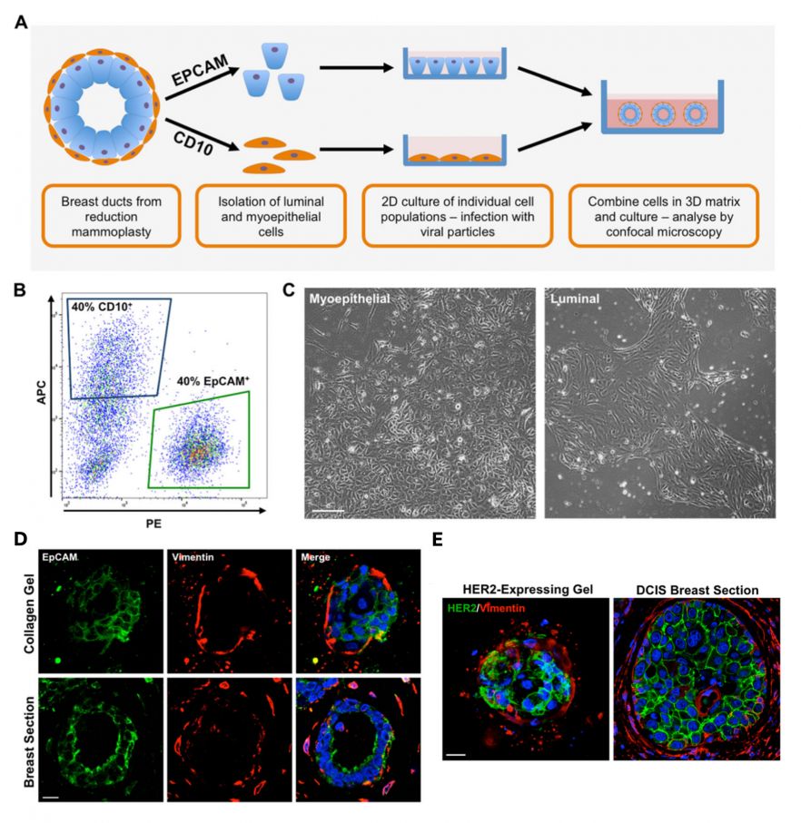
Fig. 1. Isolated myoepithelial and luminal cells maintain their characteristics in vitro. A. Schematic of proposed ductal model. B. Representative FACS plots of reduction mammoplasty specimens separated by expression of CD10 (allophycocyanin fluorescence, blue gate) and epithelial cell adhesion molecule (EpCAM; phycoerythrin fluorescence, green gate). C. Representative light micrographs of isolated myoepithelial and luminal cells grown in vitro for 10 days. Images taken at × 4 original magnification. Scale bar = 100 μm. D. Spheroid structures formed in collagen gels recapitulate a physiological breast bilayer. Expression of EpCAM and vimentin spheroid (upper) structures formed from myoepithelial and luminal cells grown in collagen for 21 days. Representative images from sections of normal human breast ducts (lower panel) are also presented. Cell nuclei are labelled with 4′,6-diamidino-2-phenylindole (blue). Scale bar = 20 μm. E. Overexpression of human epidermal growth factor receptor 2 (HER2) in the luminal compartment of a bilayer model results in destabilisation of the bilayer and luminal filling: HER2 and vimentin expression in representative HER2-expressing ductal spheroids (left) and ductal carcinoma in situ breast sections (right). Cell nuclei are labelled with DAPI (blue). Scale bar = 20 μm.
Characterisation & Validation: Extensive immunohistochemical characterisation indicates the cell types isolated represent their tissue equivalent. Comparison of structure of 3D in vitro models to tissue structures validates representation. Targeted modification of cells e.g. Her2 over-expression in luminal cells faithfully represents DCIS. Incorporation of cancer-derived fibroblasts in a ‘normal’ breast model induces a disease phenotype.
Ongoing Research: Inclusion of immune cells and adipocytes as cell types which influence cancer progression
Research Team: Louise Jones, Michael Allen, Iain Goulding
Lead Contact: Louise Jones
Last updated 06/05/2026