
Vascular Organoid
Context of Use or Disease: Vascular inflammation, vascular disease, atherosclerosis, angiogenesis, vascular calcification
DOI: Stem Cell Research & Therapy 2024, European Journal of Cell Biology 2024
Platform: Organoid
Description: Vascular organoids (VOs) were generated from human induced pluripotent stem cells (hiPSCs) in 1 % agarose-coated flat-bottomed 96-well plates. These vascular organoids can be maintained in ultra-low attachment 96-well plates with endothelial cell medium (ECM) containing 15% foetal bovine serum (FBS), 100 ng/mL VEGF, and 100 ng/ml for up to 45 days. Alternatively, they can be used to simulate multiple vascular diseases such as vascular inflammation, atherosclerosis and vascular calcification, as well as to study angiogenesis.
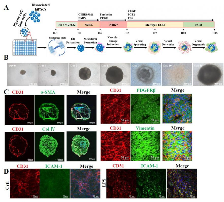
Fig. 1. Generation and characterization of VOs. A. Schematic diagram illustrating the protocol used to generate VOs from hiPSCs. ECM, endothelial cell medium. B. Bright-field images depicting the development of VOs over a 15-day differentiation period. C. Co-immunostaining of CD31 with α-SMA, PDGFRβ, Col Ⅳ, or Vimentin in D15 VOs. α-SMA, alpha- smooth muscle cell actin; Col Ⅳ, Collagen IV. D. Immunostaining of ICAM-1 expression in D15 VOs after treatment of LPS (2 μg/mL) for 24 hours.
Characterisation & Validation: Both endothelial and stromal cells within VOs show characteristic phenotypic identity, evidenced by high expression levels of CD31 for endothelial cells, and α-SMA, PDGFRβ, and vimentin for stromal cells. Incubation of VOs with lipopolysaccharide (LPS) induced vascular inflammation, as evidenced by increased expression of intercellular adhesion molecule-1 (ICAM-1).
Ongoing Research: Development of disease models for atherosclerosis and vascular calcification
Research Team: Qingzhong Xiao
Lead Contact: Qingzhong Xiao
Last updated 27/04/2026